From the joy of a first prenatal image to the decisive clarity of an emergency diagnosis, ultrasound remains one of healthcare’s most versatile and trusted tools. It transforms unseen anatomy into real-time images, enabling clinicians to act with precision and confidence.
The ultrasound probe may appear as a simple handheld device, yet it houses a complex system that blends physics, engineering, and medical science to capture images beneath the skin.
This guide explains the science within the probe, showing simulation educators how this knowledge shapes effective training scenarios. Clinicians gain a clearer view of the mechanics that support accurate scanning.
Product developers can draw insights to guide innovation, while procurement teams are better prepared to select technology that aligns with training and clinical needs.
How an Ultrasound Probe Converts Energy into Images
An ultrasound probe, also called an ultrasound transducer, is designed to convert one form of energy into another. It transforms electrical energy into sound waves and then back into electrical signals that the system processes into medical images.
Piezoelectric Crystals: The Core of an Ultrasound Transducer
Inside every probe are piezoelectric crystals, usually made from advanced ceramic materials. These crystals have a unique property:
- They generate an electrical charge when compressed or vibrated.
- They produce sound waves when a voltage is applied.
Think of a loudspeaker and a microphone working in reverse. A loudspeaker turns electrical signals into sound, while a microphone turns sound into electrical signals. In an ultrasound probe, the same piezoelectric crystals perform both functions by sending and receiving ultrasound waves.
From Electrical Pulses to High-Frequency Sound Waves
The ultrasound system sends brief, controlled electrical pulses to the piezoelectric crystals. These pulses cause rapid vibrations, creating high-frequency ultrasound waves that travel into the body.
This energy conversion is the first step in transforming invisible anatomy into visible, real-time images, forming the foundation of modern ultrasound imaging.
The Echo Principle in Ultrasound Imaging
An ultrasound probe operates through a precise cycle of actions that turn sound waves into diagnostic images. This cycle involves sending ultrasound waves into the body and converting the returning echoes into visual data.
The process follows three key steps:
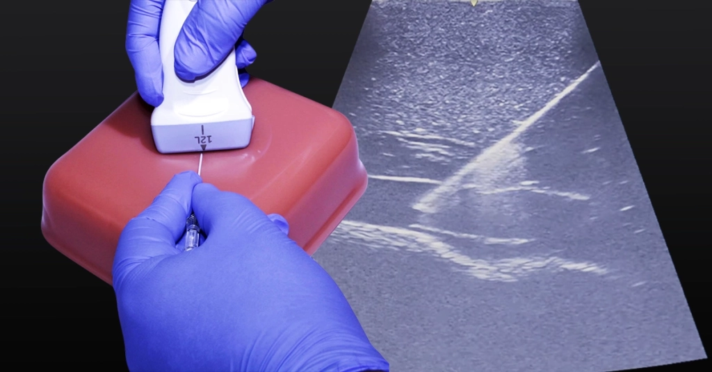
Step 1: Sending Ultrasound Waves into the Body
The sequence begins when the probe emits a short pulse of high-frequency sound waves. These waves travel through tissues and reflect off different structures, such as organs, bones, or fluid boundaries.
Step 2: Capturing the Returning Echoes
The reflections, known as echoes, return to the probe and strike the same piezoelectric crystals that generated the waves. This causes the crystals to vibrate, which converts the sound energy back into electrical signals.
The process works much like sonar in a submarine, where sound pulses are sent out and the returning echoes reveal what lies ahead.
Step 3: Converting Echo Data into Images
The ultrasound system measures how long it takes for each echo to return to determine the depth of the structure.
- Stronger echoes indicate dense or highly reflective surfaces.
- Weaker echoes suggest softer or fluid-filled areas.
Advanced algorithms then process these signals into a real-time, two-dimensional image, giving clinicians a clear view of internal anatomy for diagnosis, treatment planning, and simulation training.
Why Different Shapes Deliver Different Views
Ultrasound imaging needs vary across medical specialties. To meet these needs, manufacturers design probes with different shapes, sizes, and technologies. Each design is optimized to capture specific views and navigate different scanning environments.
One widely used design is the sector probe ultrasound, which operates through phased array technology. Its performance can be understood in three key aspects:
1. Understanding the Sector Probe Image Shape
Sector probes produce a pie-slice or fan-shaped image. This design is ideal for visualizing deep structures from a narrow point of contact on the body.
2. How Phased Array Technology Works
Inside a sector probe are multiple tiny piezoelectric elements. These elements are activated with precisely timed electrical signals, known as phase shifts. This timing steers and focuses the ultrasound beam electronically, eliminating the need to physically move the probe to change the scanning angle.
3. Key Applications of the Sector Probe
The ability to scan through a small acoustic window makes sector probe ultrasound valuable in areas with limited access, such as between ribs for cardiac imaging. They are also used in certain abdominal scans, where a wide field of view is needed from a narrow contact point.
You can learn more about specific uses on our ultrasound probe applications page.
The Impact of Ultrasound Probe Knowledge Across Healthcare Roles
A deeper grasp of ultrasound probe technology benefits different stakeholders in unique ways. Understanding how the probe works supports better training, innovation, and decision-making across healthcare and simulation.
Its impact can be seen in four key areas:
Area 1: Benefits for Simulation Educators
Knowledge of probe mechanics helps design realistic simulation scenarios, select appropriate ultrasound phantoms, and teach learners optimal probe handling and image acquisition techniques.
Area 2: Benefits for Clinicians
Clinicians who understand the probe’s function can optimize image quality, troubleshoot artifacts, and interpret scans with greater accuracy in daily practice.
Area 3: Benefits for Product Developers
For those involved in device design, this understanding guides innovation in probe construction and ensures new systems meet evolving clinical requirements.
Area 4: Benefits for Procurement Teams
Procurement specialists can make more informed equipment selections by considering probe types, features, and compatibility with training or clinical goals.
When the people using or selecting ultrasound technology understand its underlying principles, they can make better choices, improve outcomes, and ensure the right tools are in place for both training and patient care.
Better Ultrasound Training with Humimic Gels
You now understand how the probe turns energy into images. The next challenge is applying that knowledge to get clearer scans, teach with confidence, validate designs faster, and choose equipment that supports your goals.
Humimic Medical helps make that jump practical. Our ultrasound-compatible gels and phantoms enable lifelike, repeatable practice and clean testing. You also get guidance that keeps setup simple and aligns materials to your curriculum or R&D plan.
Contact us to outline a simple next step for your program.