
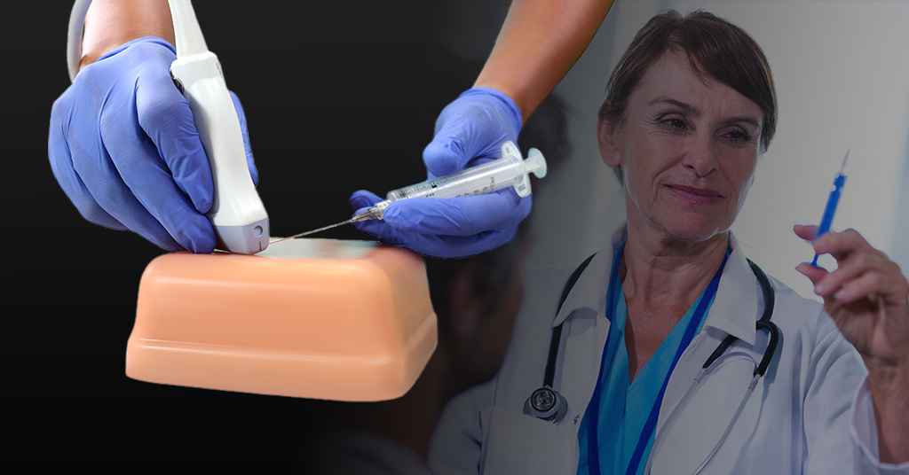
Ultrasound training often runs on tight schedules with limited patient access. Building consistent skills in probe control, needle alignment, and depth perception can be difficult.
Ultrasound phantom models are calibrated training tools that mimic how tissue looks on ultrasound and how it feels to the hand and needle. They make practice safe, repeatable, and measurable for educators, clinicians, and innovators. At Humimic Medical, our tissue-mimicking materials are shaped by field feedback and proven in high-use programs.
Evidence supports this approach. A 2024 review in BMC Medical Education found that simulation tools such as phantoms help standardize procedures and expose learners to rare findings, improving outcomes.
What’s ahead: how phantoms mimic tissue, commercial vs. DIY options, key training uses, a quick DIY recipe, and a checklist for choosing the right model.
For training value, a phantom must match what the scanner sees and what the hand feels. High-quality models achieve this through three design elements that align with ultrasound physics and procedural haptics.
The core elements are:
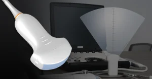
Formulations are tuned to a sound speed near 1,540 m/s, attenuation around 0.5–1.0 dB/cm/MHz, and acoustic impedance close to 1.6 MRayl. These targets support accurate depth calculation, stable beam focus, and fewer geometry-related artifacts during scanning.
Fine scatterers such as glass microspheres, cellulose fibers, or graphite create familiar speckle and contrast. Anechoic channels (typically 2–6 mm in diameter) stand in for vessels or cysts, and discrete targets enable needle-to-target visualization and landmark identification on B-mode.
Matrix stiffness is set by gel density within common Shore 00 ranges to produce predictable needle drag, insertion force, and track visibility. Consistent haptics help learners build hand–eye coordination and maintain needle alignment across sessions.
Why this matters:
These specifics let educators standardize labs, clinicians trust on-screen and tactile feedback, and developers evaluate devices under repeatable conditions without patient risk.
Choosing between commercial and DIY options depends on training goals, session volume, and how much consistency you need. The comparison below uses clear criteria so educators, clinicians, and R&D teams can match an ultrasound phantom to real program requirements.
Here’s a side-by-side comparison of ultrasound phantoms:
| Criteria | Commercial ultrasound phantoms | DIY ultrasound phantoms |
| Primary use | Standardized teaching, credentialing, skills checks, device evaluation | Intro demos, early exploration, low-frequency practice |
| Training volume | High; reliable across many sessions | Low; best for occasional use |
| Session lifespan | Months to years with routine care | Days to a few uses before degradation |
| Image consistency | Predictable texture and targets across sessions | Variable between batches |
| Needle feedback | Stable insertion feel; surfaces recover to extend use | Tracks persist and alter practice after a few passes |
| Target options | Documented vessel sizes and landmarks; repeatable layouts | Grapes, olives, balloons as simple targets |
| Maintenance | Wipe per care guide; tolerates repeated disinfection | Refrigeration required; higher contamination risk |
| Shelf life | Long when stored as directed | Short; prone to spoilage |
| Documentation | Setup, care, and performance guidance provided | None or self-created notes |
| Cost profile | Higher upfront; lower cost per session over time | Low upfront; higher cost per consistent result |
| Best for | High-volume labs, assessments, R&D workflows | Classroom demos, quick trials, target prototyping |
Commercial ultrasound phantoms are a better fit for assessments, skills checks, or device testing. Humimic Medical provides models designed for many sessions with a predictable on-screen texture and reliable needle feel.
Ultrasound phantoms help teams build skills, standardize sessions, and measure progress. Programs use them to give learners safe, repeatable practice and to create controlled test conditions for development work.
The core applications include:
Programs rely on ultrasound phantoms to reduce variability and document improvement over time. Next, see how to build a simple model for introductory practice.
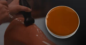
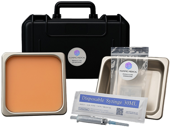
For introductory practice or quick prototyping, a simple gelatin build can demonstrate probe control, needle guidance, and target visualization. The recipe below uses common supplies and produces scan-friendly results for basic sessions.
Unlike food-grade gelatin or agar, SimuGel™ is a synthetic, reusable medium designed for medical training. It delivers calibrated ultrasound imaging, resists breakdown over repeated use, and provides a consistent platform for teaching, testing, and innovation.
DIY ultrasound phantom SimuGels are useful for foundational training and classroom demos. When programs need many sessions, stable needle feel, and documented care, teams often move to commercial ultrasound phantoms from established vendors such as Humimic Medical.
Match the ultrasound phantom to your goals, training volume, and documentation needs. The checklist below reflects what most programs evaluate before purchase.
Most product pages and datasheets include sample images by probe frequency, target dimensions, care guidance, and expected session lifespan to support side-by-side comparison; the same details are available from Humimic Medical.
Choose the option that aligns with your skill goals, throughput, and need for repeatable results. A practical rule is simple: DIY for introductory practice and concept trials, commercial for standardized teaching, assessments, and testing.
Make every training session count. Humimic Medical phantoms deliver reliable image texture, realistic needle feel, and targets designed for vascular access, biopsy, and pattern recognition. Instructors can standardize each session and easily track learner progress.
Our phantoms include exact vessel sizes, repeatable target setups, and sample images matched to common probe frequencies. Clear setup and care instructions keep your classes running smoothly and on time.
Contact us now to find the right ultrasound phantom for your equipment, training volume, and goals. Get sample images and start improving skills today.
We design trauma trainers, gel models, and procedural tools customized to your exact use case — all powered by Humimic SimuGel™.