
You’ve set up the ultrasound machine, the gel is ready, and your learner picks up the probe—only to scan the wrong plane or miss the anatomy entirely. It’s not a lack of effort. It’s often the wrong probe for the job.
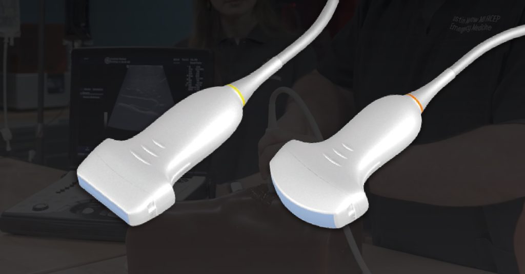
In ultrasound education, understanding the difference between a curvilinear probe and a linear probe can make or break a training session. Each one serves a specific purpose. Knowing when and why to use them helps learners gain clarity faster and scan more effectively.
In this guide, we’ll break down the differences between curvilinear and linear ultrasound probes. We’ll explore how frequency affects imaging. You’ll also learn how probe movements vary and what that means for simulation training, skill development, and patient care.
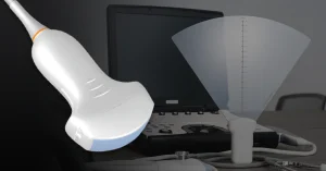
A curvilinear probe, also known as a convex probe, is designed for deep tissue imaging. It operates at low frequencies, typically between 2 to 5 MHz, which allows for greater penetration into the body but at the cost of lower image resolution 1. This makes it ideal for scanning large anatomical areas such as the abdomen, pelvis, and retroperitoneal space 2.
Due to its depth capabilities and wide field of view, the curvilinear probe is commonly used in:
Its fan-shaped beam geometry results from the curved arrangement of piezoelectric elements, producing a trapezoidal image that widens with depth3. This geometry is crucial for learners to understand, as it affects image orientation and can lead to distortion or compression if the probe is angled incorrectly.
In simulation-based education, curvilinear probes are used to teach procedural ultrasound skills. A comparative study found that curvilinear probes are just as effective as linear probes in measuring muscle mass, with intra-rater and inter-rater reliability scores above 0.95 4. This supports their use in training environments where versatility and depth imaging are required.
When instructing with a curvilinear probe:
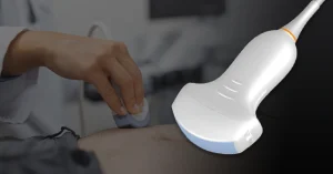
A linear ultrasound probe is designed for precision. It operates at a higher frequency, typically between 5 to 15 MHz, and is optimized for scanning structures close to the surface.
It’s commonly used for:
Here are the key characteristics:
In simulation, the linear probe helps learners practice techniques that rely on subtle adjustments in tilt and pressure. It’s essential for reinforcing hand-eye coordination and image interpretation in surface-level procedures.
Ultrasound isn’t just about the machine or the probe. It’s also about how the probe is moved. Understanding probe movements is essential for accurate imaging, and each probe type behaves differently based on its footprint and field of view.
There are four foundational ultrasound probe movements:
Why movement matters:
Help learners connect each movement to what they see on screen. Focused practice with both probe types builds confidence faster.
Each ultrasound probe is built for specific imaging tasks. Understanding when to use a curvilinear or linear probe improves scan quality, reduces setup time, and supports better clinical decision-making.
Here’s a quick comparison:
| Probe Type | Best For | Frequency | Image Detail |
| Curvilinear | Abdominal, OB/GYN, trauma, deep organs | Low (2–5 MHz) | Moderate, wide view |
| Linear | Vessels, thyroid, soft tissue, nerve blocks | High (5–15 MHz) | High, shallow structures |
Use a curvilinear probe when:
Use a linear probe when:
Tip: Start simulation sessions by explaining why you’re using a specific probe. This helps learners understand probe selection as part of clinical reasoning, not just device handling.
Choosing between a curvilinear and linear probe is more than a technical decision. It shapes how learners engage with ultrasound, how accurately they interpret what they see, and how confidently they perform procedures.
For simulation educators and clinical trainers, making probe selection part of early instruction helps build foundational skills that transfer into real-world care.
We design ultrasound training models that are reusable, adaptable, and easy to integrate into your curriculum. From probe positioning to tissue targeting, Humimic Medical tools support a wide range of training goals. See how our products fit your program today.
Most curvilinear probes operate between 2–5 MHz for deeper penetration.
The linear probe offers clearer, higher-resolution views of superficial vessels near the skin. Curvilinear is not ideal for shallow structures.
It depends. Curvilinear is best for deep organs, and linear is better for superficial structures. Using both helps trainees understand context.
With a curvilinear probe, ‘fan’ motion gives a wider field of view. Linear probes benefit from small tilting and fine adjustments.

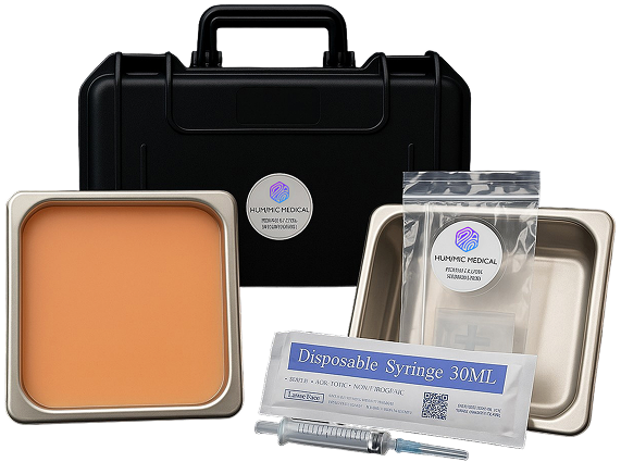
We design trauma trainers, gel models, and procedural tools customized to your exact use case — all powered by Humimic SimuGel™.