

Main takeaways:
You’re scanning for a liver mass, but your linear probe can’t penetrate deep enough. The image cuts off at 6 centimeters. The lesion sits at 12 centimeters. You adjust the gain and increase the depth setting. Still nothing but noise.
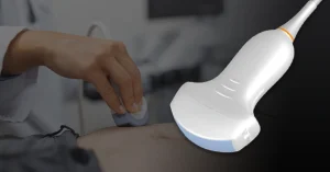
A curvilinear transducer solves this problem. The curved footprint and lower frequency waves penetrate deeper while maintaining a wide field of view. It’s built for abdominal, pelvic, and obstetric imaging where depth and coverage matter more than surface detail.
In this guide, you’ll explore what makes curvilinear transducers distinct and valuable for clinical imaging and ultrasound education.
To understand when and why to use a curvilinear transducer, we need to start with its basic structure and purpose.
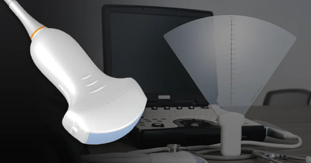
A curvilinear transducer is an ultrasound probe with a curved surface that creates a fan-shaped image. This design makes it ideal for viewing deep organs and large anatomical areas. The convex shape distinguishes it from flat linear probes and determines when and how it should be used.
Each component of the curvilinear transducer’s design plays a specific role in its performance during ultrasound scanning.
Feature 1: Curved Contact Surface
The convex footprint fits against body contours and creates a wider viewing area at depth. This design isn’t arbitrary. It’s engineered to maximize acoustic contact while giving the beam room to spread as it travels deeper.
Feature 2: Lower Frequency Range
Curvilinear probes typically operate between 2 and 5 MHz. This frequency range is optimized for deeper penetration through abdominal and pelvic tissue. Lower frequencies sacrifice some resolution but gain the depth needed to reach organs that sit 5 to 25 centimeters below the skin surface.
Feature 3: Fan-Shaped Beam Pattern
The fan shape expands the image as depth increases. This shows more anatomy in a single frame. It reduces the need for multiple probe positions during organ assessment.
This section shows how the transducer’s internal mechanics shape the visual output and affect real-world scan performance.
The transducer sends ultrasound waves into tissue. As the beam travels, it spreads outward in a fan pattern. Reflected echoes return to the probe and are processed into an image on screen.
The wider footprint at depth helps visualize large organs like the liver, kidneys, or uterus without repositioning the probe repeatedly. This makes curvilinear transducers efficient for abdominal, obstetric, and pelvic exams.
Understanding these design principles helps educators select the right training tools. It also helps learners recognize when to reach for a curvilinear probe instead of a linear one.
Both tools are ultrasound transducers, but they serve different imaging goals. Here’s how they compare based on design, application, and performance.
Here’s how the two probe types compare across clinical scenarios. Use this comparison to decide which probe to pick depending on your clinical task:
The main difference is shape and purpose. A curvilinear transducer has a curved footprint for deep, wide-view imaging. A linear ultrasound probe has a flat footprint for high-resolution imaging of superficial structures.
Each probe type serves different clinical needs based on target depth and required image detail. Choosing between them depends on the anatomy you’re scanning and how deep the target is.
|
Criteria |
Curvilinear Transducer |
Linear Ultrasound Probe |
| Footprint shape | Convex (curved) | Flat (rectangular) |
| Frequency range | 2–5 MHz | 5–15 MHz |
| Image shape | Fan-shaped (wider at depth) | Rectangular (parallel lines) |
| Field of view | Wide, expanding with depth | Narrow, consistent width |
| Penetration depth | Deep (5–25 cm) | Shallow (1–6 cm) |
| Best for | Abdominal, obstetric, pelvic imaging | Vascular access, superficial structures, MSK |
| Common uses | Liver, kidneys, bladder, pregnancy scans | IV access, nerve blocks, tendons, thyroid |
| Image resolution | Lower resolution, better depth | Higher resolution, better detail at shallow depth |
These are the most common clinical scenarios for each probe type:
Choose curvilinear transducers when:
✓ Abdominal organ assessment
✓ Obstetric and gynecologic exams
✓ Pelvic imaging
✓ Evaluating deep vascular structures
✓ FAST exams in trauma settings
Choose linear ultrasound probes when:
✓ Vascular access procedures like IV and central line placement
✓ Nerve blocks and regional anesthesia
✓ Musculoskeletal imaging
✓ Thyroid and superficial mass evaluation
✓ Foreign body localization
When it comes to high-volume imaging or deeper tissue assessments, curvilinear probes consistently deliver. Here’s where they work best.
The curvilinear transducer is the standard choice for visualizing abdominal organs. Its lower frequency and convex shape work together to penetrate through multiple tissue layers while maintaining a wide enough view to see organ margins and surrounding structures.
Curvilinear probes are essential for pregnancy monitoring and pelvic imaging. The lower frequency range provides clear images through maternal tissue layers without compromising fetal visualization.
Focused Assessment with Sonography for Trauma (FAST) relies on curvilinear transducers to quickly detect free fluid in the abdomen and pelvis. Speed and coverage matter more than fine detail in these time-critical assessments.
Transabdominal pelvic scans require both depth and a wide view to visualize the bladder and surrounding pelvic organs without invasive endocavitary scanning.
While linear probes handle superficial vessels, curvilinear transducers are better suited for deep vascular structures that require penetration beyond 5 centimeters.
Effective ultrasound education relies on simulation tools that feel and function like real tissue. This section outlines the essentials for training with confidence.
To make practice meaningful and repeatable, your training tool needs to meet four technical criteria:
Criteria #1: Proper Acoustic Properties- Matched to soft tissue with correct speed of sound and attenuation rates.
Criteria #2: Realistic Image Texture- Fine scatterers for speckle and contrast that mimic organ parenchyma.
Criteria #3: Clinically Relevant Target Depths- Targets placed between 5–15 cm to reflect real scanning conditions.
Criteria #4: Consistent Tactile Feedback- Gel density that supports probe pressure and scanning technique without fatigue.
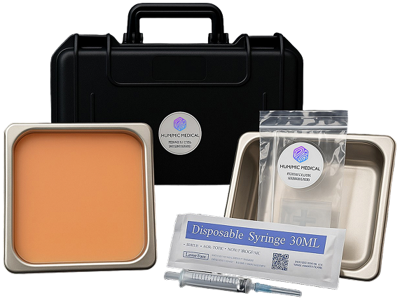
Simulation only works if it feels real. That’s why Humimic uses SimuGel™ for its ultrasound phantoms. Here’s how it supports training programs.
Below are four training scenarios where curvilinear probes paired with SimuGel-based phantoms make the biggest impact:
Scenario #1: Abdominal Scanning Practice- Learn organ identification and depth control.
Scenario #1: FAST Exam Drills- Develop speed and technique for trauma protocols.
Scenario #2: Obstetric Imaging Training- Practice fetal position and biometry without patient risk.
Scenario #4: Pelvic Imaging Sessions- Improve skills in bladder volume and pelvic mass assessment.
The probe you select can make or break the clarity of your scan. Let’s break down when to reach for each option.
Use this quick decision checklist to guide probe selection based on clinical depth, anatomy, and diagnostic need:
Tip 1: Choose curvilinear transducers when:
Tip 2: Choose linear ultrasound probes when:
Clinical learners aren’t just memorizing probe types. They’re building habits that affect real patient care. When simulation tools don’t behave like real tissue, that gap becomes visible the moment a learner picks up a probe in a live setting.
At Humimic SimuGel, we know how frustrating that disconnect can be, especially for programs trying to set students up for success from day one.
That’s where SimuGel™ comes in. Our ultrasound phantoms are designed to replicate how tissue feels, responds, and appears under ultrasound. With durability that holds up through hundreds of scans, this helps programs provide consistent, hands-on training across disciplines like abdominal scanning, FAST exams, and OB imaging.
Add a Humimic phantom to your cart and give your learners the realism they’ll trust when it counts.
Even with hands-on practice, some questions come up again and again. Here are quick answers to what many learners and educators ask about curvilinear probes.
A curvilinear probe has a convex footprint and produces a fan-shaped image ideal for deep tissue imaging (5–25 cm). A linear ultrasound probe has a flat footprint and produces a rectangular image best for superficial structures (1–6 cm) and high-resolution scanning.
Most curvilinear ultrasound probes operate between 2 and 5 MHz. This allows for deeper penetration through abdominal and pelvic tissue. Lower frequencies sacrifice some resolution but gain the depth needed for organ imaging.
Curvilinear probes can image deep vessels like the abdominal aorta or IVC. But linear probes are better for superficial vascular access procedures like IV insertion or central line placement due to higher resolution and flat footprint design.
The convex shape of the transducer sends ultrasound beams outward in a fan pattern. As the beams travel deeper, they spread apart, creating a wider field of view at increased depth. This is a physics consequence of the curved crystal array.
Phantoms designed for curvilinear training should have acoustic properties matched to soft tissue, deep target placement (5–15 cm), and consistent image texture. Humimic Medical phantoms are built with SimuGel to provide realistic imaging and repeatable results for curvilinear and linear probes across multiple training sessions.

We design trauma trainers, gel models, and procedural tools customized to your exact use case — all powered by Humimic SimuGel™.