

Main points:
Curvilinear ultrasound probes are the preferred choice for abdominal scanning because they provide greater depth penetration, a wider field of view, and better contact with the curved abdomen surface.
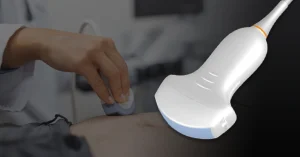
You position the probe on your patient’s abdomen and begin scanning. The image appears grainy and incomplete. You can’t capture deep structures clearly. The problem isn’t your technique. It’s your probe selection.
A 2024 clinical evaluation found that using a transducer designed for deep abdominal imaging improved B‑mode image quality by up to 53% and color Doppler performance by up to 73%, demonstrating how the right probe can significantly increase diagnostic clarity. For abdominal scanning, that choice is almost always curvilinear.
In this article, we’ll explain what makes curvilinear probes essential for abdominal imaging, how they differ from other probe types, and how to train effectively with them using realistic simulation.
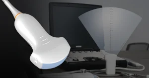
A curvilinear probe (sometimes called a convex or curved array probe) is an ultrasound transducer with a rounded, convex surface that emits sound waves in a fan-shaped pattern. This design is fundamentally different from linear probes, which have flat surfaces and produce rectangular images.
Curvilinear probes operate at lower frequencies (typically 2-5 MHz) than linear probes, which typically use 7-15 MHz. This lower frequency allows the sound waves to penetrate deeper into the body. This feature becomes essential when imaging abdominal organs that may be 10-15 cm beneath the skin surface.
Think of the difference like this:
This trade-off between depth and resolution explains why curvilinear probes are the standard choice for abdominal imaging across major clinical guidelines, including FAST, aortic, biliary, renal, and pelvic exams.
Three key advantages make curvilinear probes ideal for abdominal scanning:
Abdominal organs aren’t superficial. The aorta, kidneys, and portions of the liver can be 8-15 cm from the skin surface. The lower frequency of curvilinear probes allows sound waves to penetrate this distance without losing too much signal strength.
When using a curvilinear probe:
By contrast, linear high-frequency probes typically max out at 5-7 cm depth. This works fine for vascular access but becomes insufficient for most abdominal structures.
The fan-shaped beam from a curvilinear probe creates a field of view that expands with depth. This typically spans 60-70 degrees wide. This means:
This wider perspective reduces the number of probe adjustments needed. The result is more efficient and comprehensive exams.
The curved face of a curvilinear probe fits naturally against the convex surface of the abdomen. This improved contact:
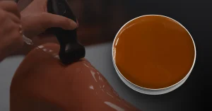
The importance of this anatomical match shouldn’t be underestimated. Proper probe-to-skin contact is essential because ultrasound waves cannot travel through air; coupling gel eliminates air gaps and ensures accurate image transmission.
Despite their advantages for abdominal imaging, curvilinear probes present several training challenges:
Unlike linear probes that create rectangular images, curvilinear probes produce images that widen with depth. This fan-shaped perspective requires trainees to:
Mastering this visual adjustment takes practice, but it’s a key step toward accurate interpretation. A 2023 study by Walsh et al. found that novices had 5.4% higher accuracy with curvilinear images than with phased array images—showing the unique learning curve and value of understanding fan-shaped views.
The deeper imaging of abdominal scans requires careful adjustment of:
Deeper imaging adds another layer of complexity. Abdominal ultrasound typically requires 8–10 cm depth in thin patients and up to 25–30 cm in obese patients to visualize key structures such as the aorta and liver. This range highlights why fine-tuning parameters like depth, gain, and focal zones is essential for clear, diagnostic images.
Effective abdominal scanning requires specific handling techniques:
These skills take time and guided repetition to develop. Educational guidelines and studies show that achieving basic competency in abdominal ultrasound scanning, especially with curvilinear probes, requires around 25–30 hours of supervised hands-on practice.
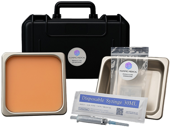
Training effectively with curvilinear probes requires a medium that replicates human tissue acoustically while providing consistent, repeatable practice opportunities. This is where Humimic SimuGel™ makes a significant difference.
Humimic SimuGel™ is specifically engineered to mimic the acoustic properties of human tissue, creating ultrasound images that closely resemble clinical scans. The crystal-clear material provides ideal conditions for visualizing simulated structures at various depths.
SimuGel ultrasound phantoms offer several advantages specifically for curvilinear probe training:
For curvilinear probe training, educators typically select:
Each technique helps trainees build spatial awareness, interpret real-time images, and apply proper probe control. These fundamentals form the basis of accurate ultrasound scanning.
When training with SimuGel phantoms, instructors emphasize five core techniques that transfer directly to clinical practice:
Maintaining continuous contact between the curved probe surface and the scanning surface is critical. Trainees learn to:
Efficient abdominal imaging follows organized patterns:
“The SimuGel’s clear consistency lets students see their probe position relative to embedded structures,” notes clinical instructor Jamie Roberts. “This visual feedback helps them connect hand movements to image changes.”
Setting appropriate depth is crucial for balancing penetration and frame rate:
Proper gain settings compensate for attenuation at different depths:
Strategic focal zone positioning enhances resolution:
The skills developed through practice with curvilinear probes on SimuGel phantoms translate directly to a wide range of clinical scenarios:
A recent study found that point-of-care ultrasound (POCUS) for suspected small bowel obstruction reduced median time to diagnosis from 217 minutes to 121 minutes compared to standard imaging workflows.
Getting consistent results can be a challenge. We understand how unpredictable materials and tools can affect your work and slow down progress.
Humimic Medical is here to make things more reliable. Our solutions are designed to support professionals who need precision, consistency, and confidence in every task.
Contact us today to find a dependable partner for your next project.
Most adult abdominal imaging is performed in the 3.5-5 MHz range. This provides an optimal balance between penetration and resolution for average adult tissue depth.
Curvilinear probes are not optimal for superficial structures. Linear high-frequency probes (7-15 MHz) provide better resolution for structures less than 5 cm deep.
SimuGel offers comparable acoustic properties to commercial phantoms but with greater durability through thousands of scans. Unlike gelatin-based models that deteriorate over time, SimuGel maintains consistent performance through repeated use.
SimuGel’s consistent density and acoustic properties closely mimic human tissue while allowing for visual verification of target structures. This combination creates an ideal learning environment that bridges demonstration and clinical practice.
We design trauma trainers, gel models, and procedural tools customized to your exact use case — all powered by Humimic SimuGel™.